Sunday, 24 December 2017

Enzymes (Basics)

Enzymes (Basics)

Enzymes are biological catalysts and help speed up reactions, many globular proteins are enzymes.


Metabolism which is the sum of all the chemical reactions occurring in a an organisms. Enzymes control these metabolic reactions which can either be:


-Anabolic = The formation of molecules from smaller units.


-Catabolic= The breaking down of larger molecules.


Activation energy is the amount of energy that is needed for a reaction to start. 


An example of this could be seen in my previous post where I talked about DNA replication and uses the enzyme DNA Polymerase.


Enzymes can intracellular (work inside cells) or extracellular (work outside cells) like some bacteria secrete digestive enzymes into the environment to digest their food before consumption.


1-Substrates collide with the active site of the enzyme.2-The shape of the active site is complementary to the substrate.3-The substrate bids to the active site to form an enzyme-substrate complex (ESC)4-Bonds in the substrate are placed under strain and break the enzyme provides an alternative reaction pathway that reduces the activation energy required for the reaction.5-An enzyme product complex is produced and the products can be released.


The lock-key hypothesis:That the shape of the active site is a complementary site for the substrate molecule and is therefore specific to one substrate.


The Induced fit hypothesis:

When the substrate molecule binds to the active site it initially has a weak binding by the substrate will alter the enzyme structure, this strengthens the temporary bonds between the substrate and the enzyme and weakens the bonds within the substrate.

**REMEMBER TO STAY POSITIVE LIKE A PROTON!!**

Monday, 18 December 2017

DNA Replication


 DNA Replication

DNA Replication results in two identical double helices where genetic information is conserved in each helix and is semi conservative as one strand is new and one strand is from the original strand.

The genetic code uses a triplet code which is called a codon and codes for the production of one amino acid.  The code is degenerate so means one amino acids can have many different types of cordons for the same amino acid.

1-DNA unzips by helicase enzyme to create replication fork and both DNA strands act as a template.

2-Primase enzyme makes a small piece of RNA called primer.

3-DNA polymerase binds to primer and adds bases to 5'-3' end and the leading strand is made continuously 5'-3' end.

Free monomer nucleotides are activated by phosphate groups are added to them and free nucleotides form hydrogen bonds with base pairs (A=T) purines and (C=G) Pyrimidines.
as lagging strand is 3'-5' end primase makes primers every 120 bases to create Okazaki fragments.

4-Exonuclease remove DNA primers.

5-DNA polymerase fills any gaps left over.

6-DNA ligase seals to create a continuous strand.


**REMEMBER TO STAY POSITIVE LIKE A PROTON!!**

Sunday, 10 December 2017

Cell signalling G proteins

Cell signalling G proteins

Cell signalling is a type of communication that cells do with one another or with itself. It involves transmitting ligands which are signalling molecules which then bind to a receptor protein to create a change such a release a chemical or hormone.

G-Proteins guanine-nucleotide binding protein.

G-Proteins coupled receptor structure:
-7 transmembrane alpha helices with a extracellular N terminus and a cytosolic C terminus containing a ligland (signalling molecules) binding site also there is a cytosolic loop between segment 5 and 6 binds specific G proteins.

GPCR (G protein coupled receptors) are very common types of receptors on the plasma membrane and most drugs produced work around these types of receptors. 

1-Many G protein coupled receptors have a large extracellular ligland binding domain when an appropriate  protein ligland binds to this domain the receptor undergoes a conformation change that is transmitted to its cytosolic regions which now activate a trimeric GTP binding protein also called g protein.

2-A G protein consists of 3 protein sub units; Alpha beta and Gamma. Alpha and Gamma have contently attached lipid tails which help anchor the G protein to the membrane, In the absence of a signal the alpha sub unit has a GDP bound to it and the G protein is inactive .

3-An activated receptor induces a conformational change in  the alpha subunit causing the GDP to dissociate and GTP in the cytsol binds in its place which  causes a conformational change activating both alpha sub unit and beta gamma complex now it can regulate the activity of target proteins in the plasma membrane

4-The activated target proteins then relay the signal to other components in the signalling cascade eventually the alpha sub unit hydrolyses its bound GTP  to GDP which inactivates the sub unit. This is sped up by a protein called RGS (regulator of g protein signalling). The inactivated alpha sub unit reforms with the beta and gamma sub units which turns off other downstream events.

5-As long as the signalling receptor remains stimulated it can continue to activate G proteins but after a very long time it begins to get inactive. so a receptor kinase phosphorylates the cytosolic portions of the activated receptor and then binds to a high affinity to arrestin protein which inactivates the receptor by preventing any interactions with G proteins.


**REMEMBER TO STAY POSITIVE LIKE A PROTON!!**

Sunday, 3 December 2017

The sliding filament model

The sliding filament model

A muscle is an example of an effectors and impulses are transmitted by motor neurons which stimulate muscle cells to contract and produce a response.  

There are different types of muscles the first one is called skeletal muscle it has a striated and striped appearance and can be attached to bones via tendons. The type of contraction is voluntary and is very fast but is short is in duration. The next is cardiac muscle it is also striated and is used in the heart and has a involuntary contraction with a intermediate speed and duration. The next type of muscle is smooth muscle and is non striated and is used in the blood vessels and digestive system and is involuntary and is slow and can be long lasting.

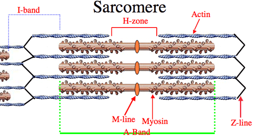
Muscle cells fuse to form fibers with each muscle fiber contains many myofibrils and are organelles made principally of having two proteins called actin and myosin. The myofibrils are then composed of many repeating units called sarcomeres.


A neuromuscular junction is the synapse between a  motor neuron and a muscle fibre and it works having the principles as a synapse between two neurons and a neurotransmitter called acetylcholine this diffuses across the synaptic cleft and binds to receptors on the sarcolemma which results in the depolarisation and a motor unit comprises all the muscles fibres which is supplied by one motor neurone.

The sliding filament model process

1-The sarcolemma is depolarised

2-The depolarisation spreads through T-tubles to sarcoplasmic reticulum a specialised smooth endoplasmic reticulum.

3-Calcium ions are released from sarcoplasmic reticulum.

4-Calcium ions binds to troponin which is a protein that is attached to actin.

5-The troponin changes shape which causes tropomysoin to be moved away from the myosin binding site it had been covering.

6-Myosin heads bind to the binding site on actin this forms cross bridges.

7-Myosin heads tilt then moves the actin this is called the power stroke and ADP is released from myosin at this stage.

8-ATP binds to mysoin causing it to detach from the actin.


9-ATP is hydrolysed to ADP causing the myosin head to resume its original position the head is free to attach further down the actin and more than 100 power strokes can be performed by each mysoin each second. 

**REMEMBER TO STAY POSITIVE LIKE A PROTON!!**